Medical Ultrasound Imaging
Saturday, 27 April 2024
Info Sheets Out- side     | 'Contrast Resolution' Searchterm 'Contrast Resolution' found in 23 articles 7 definitions [ • ] - 16 booleans [• ]Result Pages : • 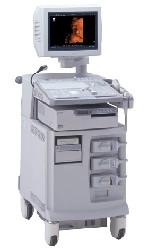 From ALOKA Co., Ltd.;
'The ProSound SSD-4000 utilizes the most advanced acoustic technologies available today, and its multidisciplinary technology architecture enables it to offer great versatility and flexibility over a wide range of clinical applications. With its new-generation, front-end technology including a 12-bit A/D converter, the ProSound SSD-4000 offers superior contrast resolution−especially when compared to 10-bit systems.'
Device Information and Specification
APPLICATIONS
CONFIGURATION
Compact, portable, dual dynamic display
Wide-band super high-density (W-SHD) transducers
OPTIONAL PACKAGE
Volume Mode
STORAGE, CONNECTIVITY, OS
Data Management Subsystem (iDMS), DICOM-Worklist
DATA PROCESSING
•
The wider the ultrasound beam, the more severe the problem with volume averaging and the beam-width artifact, to avoid this, the ultrasound beam can be shaped with lenses.
Different possibilities to focus the beam:
•
Mechanical focusing is performed by placing an acoustic lens on the surface of the transducer or using a transducer with a concave face.
•
Electronic focusing uses multiple phased array (annular or linear) elements, sequentially fired to focus the beam.
Conventional multi-element transducers are electronically focused in order to minimize beam width. This transducer type can be focused electronically only along the long axis of the probe where there are multiple elements, along the short axis (elevation axis) are conventional transducers only one element wide. Electronic focusing in any axis requires multiple transducer elements arrayed along that axis. Short axis focusing of conventional multi-element transducers requires an acoustic lens which has a fixed focal length. For operation at frequencies at or even above 10 MHz, quantization noise reduces contrast resolution. Digital beamforming gives better control over time delay quantization errors. In digital beamformers the delay accuracy is improved, thus allowing higher frequency operation. In analog beamformers, delay accuracy is in the order of 20 ns. Phased beamformers are suitable to handle linear phased arrays and are used for sector formats such as required in cardiography to improve image quality. Beamforming in ultrasound instruments for medical imaging uses analog delay lines. The signal from each individual element is delayed in order to steer the beam in the desired direction and focuses the beam. The receive beamformer tracks the depth and focuses the receive beam as the depth increases for each transmitted pulse. The receive aperture increase with depth. The lateral resolution is constant with depth, and decreases the sensitivity to aberrations in the imaged tissue. A requirement for dynamic control of the used elements is given. Since often a weighting function (apodization) is used for side lobe reduction, the element weights also have to be dynamically updated with depth. See also Huygens Principle. •
Breast ultrasound (sonography or ultrasonography) it is an important tool in the characterization of breast lesions, detected with mammography or clinical breast examination. However, a breast sonogram is not approved by the U.S. Food and Drug Administration (FDA) as a screening tool for breast cancer and is used additional to a mammogram. Ultrasound is useful in guiding needles for fine needle aspiration and core biopsies. Breast ultrasound has optimal contrast resolution, but it lacks the spatial resolution of conventional mammography and cannot provide as much detail as a mammogram image. In addition, ultrasound is unable to show tiny calcium deposits (microcalcifications) that are often early indications of breast cancer. See also Biopsy, Interventional Ultrasound, Ultrasound Safety, Side Effect and Ultrasound Regulations. • 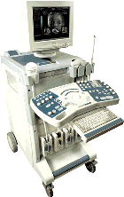 From Medison Co.,Ltd.;
'The SONOACE 9900 offers exceptional B/W image clarity by newly developed FINE™ Filter technology, which contributes to edge enhancement and average filtering in real-time. OSIO™(Organ Specified Image Optimization)sets the best diagnosis environment with minimum manual operation and increases patient throughput. Also, color images are improved with better color Doppler performance, high color sensitivity, fine pixel image, and reduced flash artifact across all probes.' 'The SONOACE 9900 PRIME from Medison offers full range of applications specially designed for your practice. With the addition of the new C-Square Processing and PSAD Beamformer, SONOACE 9900 PRIME provides higher contrast resolution and clearer image quality in all application ranges. Based on super clear image resolution and versatile 3D functions, SONOACE 9900 PRIME offers you the true value of future ultrasound technology.' •
Targeted ultrasound contrast agents provide advantages compared with usual microbubble blood pool agents. The goal of targeted ultrasound contrast agents is to significantly and selectively enhance the detection of a targeted vascular site. Tissue-specific ultrasound contrast agents improve the image contrast resolution through differential uptake. Targeted drug delivery via contrast microbubbles is another contrast media concept and provides the potential for earlier detection and characterization of disease. Targeted contrast imaging provides a higher sensitivity and specificity than obtained with a nontargeted contrast agent. The detection of disease-indicative molecular signatures may allow early assessment of pathology on a molecular level. Molecular imaging should be an efficient and less invasive technique to obtain three-dimensional localization of pathology. Ultrasound agents typically remain within the vascular space, and therefore possible targets include molecular markers on thrombus, endothelial cells, and leukocytes. Targeted contrast agents permit noninvasive detection of thrombus, cancer, inflammation, or other sites where specific integrins or other adhesion molecules are expressed. Adhesion molecules such as monoclonal antibodies, peptides, asialoglycoproteins, or polysaccharides are incorporated into the shell of the microbubble or liposome. After injection into the bloodstream, the targeted agent accumulates via adhesion receptors at the affected site, enhancing detection with an ultrasound system. See also Acoustically Active Lipospheres, and Tissue-Specific Ultrasound Contrast Agent. Further Reading: News & More:
Result Pages : | Share This Page Look Ups |
Medical-Ultrasound-Imaging.com
former US-TIP.com
Member of SoftWays' Medical Imaging Group - MR-TIP • Radiology TIP • Medical-Ultrasound-Imaging
Copyright © 2008 - 2024 SoftWays. All rights reserved.
Terms of Use | Privacy Policy | Advertise With Us
former US-TIP.com
Member of SoftWays' Medical Imaging Group - MR-TIP • Radiology TIP • Medical-Ultrasound-Imaging
Copyright © 2008 - 2024 SoftWays. All rights reserved.
Terms of Use | Privacy Policy | Advertise With Us
[last update: 2023-11-06 01:42:00]